The National University of Córdoba already has a device that allows it to analyze biological material with optical “super resolution.”
It is used for a technique called stimulation emission depletion (STED) microscopy, thanks to which you can see in detail internal structures of a bacteria such as Escherichia coli.
Due to the sophisticated way in which its lasers operate, this highly complex scientific technology focuses on samples that have a fluorescent molecule that restrict the area to be observed and achieves a resolution up to six times greater than conventional confocal microscopy, reaching up to make particles up to 80 nanometers in diameter visible in detail.
To understand what this means, you have to think that a human hair has a thickness of approximately 60,000 nanometers (nm).
The device that allows this visual range required an investment of close to 250 thousand euros, and was installed in September 2023 at the Córdoba Micro and Nanoscopy Center (Ceminco), which depends on three centers of the UNC and Conicet: the Córdoba Biological Chemistry Research Center (Ciquibic); the Mercedes and Martín Ferreyra Institute (IMMF-Conicet) and the Research Center in Clinical Biochemistry and Immunology (Cibici-Conicet).
The nano world
“This leap in optical quality allows us to observe structures at the subcellular level with unprecedented detail, revealing aspects of biology that were previously hidden from our sight,” Carlos Mas, member of Ceminco, from where this instrument is operated, explained to UNCiencia. last generation.
“For research on biological phenomena, the smaller you see, the more detail and information you obtain,” Mas clarifies. “STED microscopy offers more than six times greater optical resolution than confocal microscopy.”
And he elaborates: “The light used in optical microscopes, including confocal ones, cannot be focused beyond a certain size due to its wave nature, which causes diffraction and limits the ability to analyze small structures and details in the observed samples. . Therefore, until now, it was not possible to observe objects smaller than the wavelength used for observation. But the STED microscope overcomes this diffraction barrier, allowing tiny objects to be observed.”
As seen in the image above (1), taken with the use of STED, there is the nucleus of a cell, which thanks to the florophores molecules that serve as “highlighters” that mark the DNA, the genetic material of the cell, and other structures.
It is a cell grown in a laboratory, which has the protein actin “painted” in red, one of the most important in the cellular skeleton and what gives it its shape. Another protein located in the Golgi Complex is seen in green; and in magenta another protein of the nuclear pore complex, which acts as a gate that regulates the passage of proteins, RNA and other molecules, and that facilitates basic processes for life, such as gene translation.
“In this image – comments the Ceminco specialist – you can see the area with a white box that indicates the location where an observation was made with STED microscopy (Photo below) which in turn reveals more precise structural details that cannot be detected with confocal microscopy.
In image 2 you can see another photo of a cell and the comparison of the same area with confocal microscopy, which shows the level of detail achieved with this new technology.
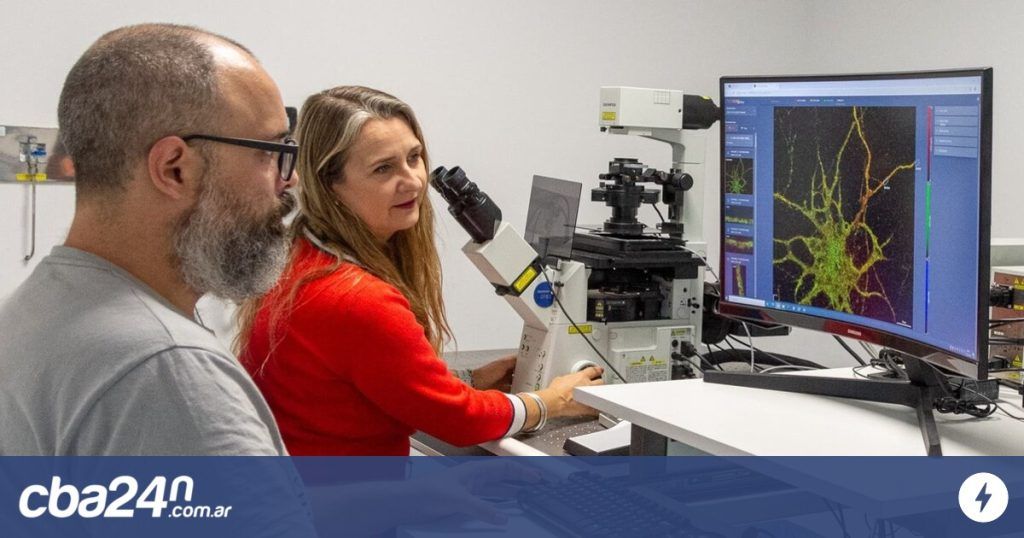
Another use case of STED microscopy in Córdoba is image 3, which shows axons, the “wires” that communicate with neurons in research that attempts to describe the causes of spinal muscular atrophy.
STED microscopy was also used to study “nanovesicles” (with sizes between 80 and 250 nm) released by stem cells in humans (image 4). In this case, the aim was to explore applications in regenerative medicine and how these structures can serve as biomarkers in diseases that affect the nervous system.
A sea of applications
According to Ceminco, this type of microscopy allowed crucial advances in the world in the understanding of cellular processes such as endocytosis, exocytosis and intracellular signaling.
In biomedicine, STED microscopy is used to study neurodegenerative diseases, cancer, viral infections and other pathologies, providing valuable information for the creation of new therapies.
All work linked to nanosciences demands this technology, such as the characterization of nanostructures, the distribution of nanoparticles or the study of interaction at the nanometric scale.
Meanwhile, in industry, STED microscopy is used for quality control of pharmaceutical products, medical devices and high-tech electronic components.
Ceminco offers this STED microscope to the entire scientific community and the private sector.
Cost. It rents the use of STED microscopy in exchange for very affordable rates, which start from $16 (official dollar) per hour for UNC users to $32 for the private sector.
Consultations. Contact Ceminco from Monday to Friday from 9:30 a.m. to 6:30 p.m. at 0351-5353735 Ext. 90503/2.
Publication date: June 20, 2024