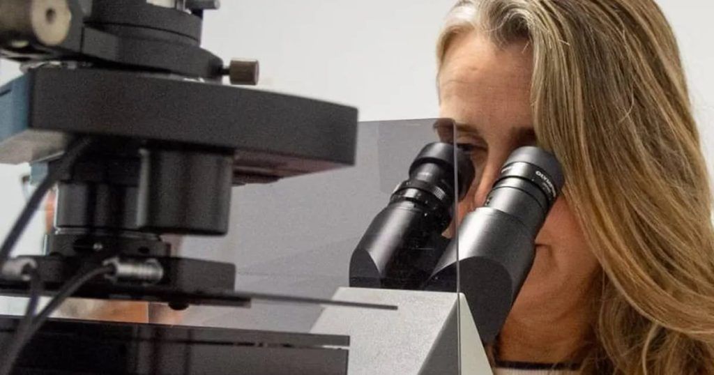
The National University of Córdoba (UNC) has a microscope that will allow biological material to be analyzed with optical “super resolution.” It is the only one in Córdoba and one of the few nationwide.
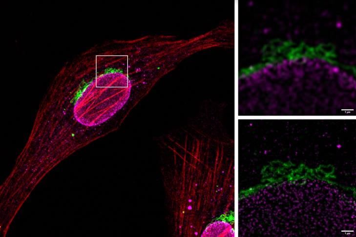
The device uses a technique called stimulation emission depletion (Sted) microscopy, through which the internal structures of a bacteria such as Escherichia coli can be seen in detail.
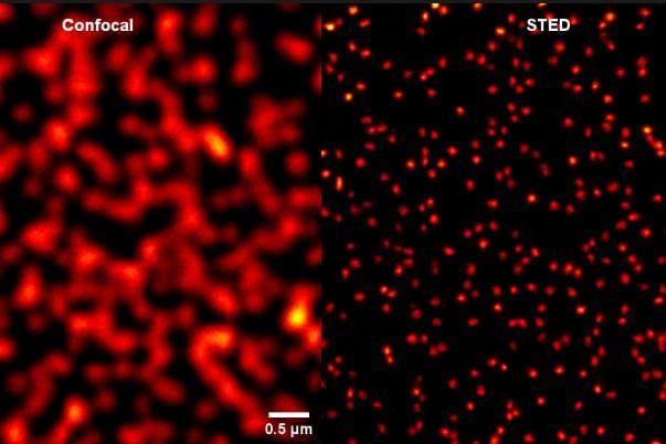
Due to the sophisticated way in which its lasers operate, this highly complex scientific technology focuses on samples that have a fluorescent molecule that restricts the area to be observed. And they achieve a resolution up to six times greater than conventional confocal microscopy, being able to make particles up to 80 nanometers in diameter visible in detail.
To understand what it means, you have to think that a human hair has a thickness of approximately 60,000 nanometers (nm).
This super microscope required an investment of close to 250 thousand euros, and was installed in September 2023 at the Córdoba Micro and Nanoscopy Center (Ceminco), which depends on the UNC and Conicet.
“It is a microscope that works with florescence, in most research they already use it and it allows them to be familiar with fluorescent markings. Only this one works with special fluorescent molecules that are resistant to photobleaching (property of erasing fluorescence),” he told The voice Carlos Mas, member of Ceminco, from where this state-of-the-art instrument is operated.
The super microscope will benefit most of the life sciences research lines in Córdoba, and the rest of the country. “It will allow them to study with much more resolution details than other types of microscopes, from a 10X lens to a 60X lens,” he stated.
Furthermore, he highlighted that any researcher, from any part of Argentina, will be able to have access to this technology since Ceminco is a member of the national microscopy system.
And he added: “At the research level it is a very important leap in technology and the effect will be seen in a few years. Now, researchers will be able to think about projects where the answers come from super resolution.”
The nano world
Mas said that “this leap in optical quality allows us to observe structures at the sub-cellular level in unprecedented detail, revealing aspects of biology that were previously hidden from our view.”
Furthermore, he expressed that for research on biological phenomena, “the smaller you see, the more details and information you obtain. “Sted microscopy offers more than six times greater optical resolution than confocal microscopy.”
And he highlighted that the light used in optical microscopes, including confocal ones, cannot be focused beyond a certain size due to its wave nature, which causes diffraction and limits the ability to analyze structures and small details in the samples observed. .
Therefore, “until now, it was not possible to observe objects smaller than the wavelength used for observation, but the Sted microscope overcomes this diffraction barrier, allowing tiny objects to be observed.”
Various applications
From Ceminco, they stated that this type of microscopy allowed, worldwide, crucial advances in the understanding of cellular processes such as endocytosis, exocytosis and intracellular signaling.
In biomedicine, Sted microscopy is used to study neurodegenerative diseases, cancer, viral infections and other pathologies, providing valuable information for the creation of new therapies.
All work linked to nanosciences demands this technology, such as the characterization of nanostructures, the distribution of nanoparticles or the study of interaction at the nanometric scale.
Meanwhile, in industry, it is used for quality control of pharmaceutical products, medical devices and high-tech electronic components.
Where to rent it
Ceminco offers the Sted microscope to the entire scientific community and the private sector. The rental cost for its use starts at $16 per hour (official dollar) for UNC users, and up to $32 for the private sector.
Consultations: Monday to Friday from 9.30 a.m. to 6.30 p.m. at 0351-5353735 extension 90503/2.