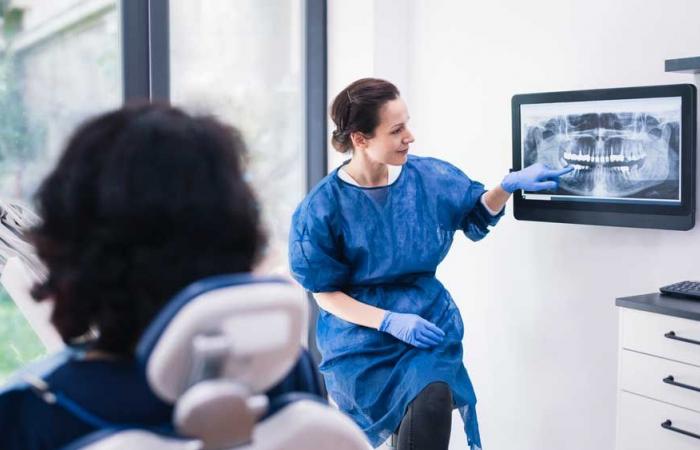
Early detection and timely treatment is important for the success of dental implant treatment.
Summary
The peri-implant diseases are common complications that occur around osseointegrated endosseous implants and are the result of an imbalance between the bacterial challenge and the host response. Peri-implant diseases can only affect the peri-implant mucosa (peri-implant mucositis) or also affect the supporting bone (peri-implantitis). Early detection of peri-implant diseases and timely treatment is important for the success of dental implant treatment. He peri-implant probing It is essential to evaluate the peri-implant health status and should be performed at each control visit. Dentists must be familiar with the clinical and radiological characteristics of both conditions in order to make an accurate diagnosis and determine the appropriate treatment required. This article aims to provide clinicians with an understanding of the key differences between peri-implant health, peri-implant mucositis, and peri-implantitis.
| Key points
|
Introduction
Peri-implant diseases, peri-implant mucositis and peri-implantitis refer to inflammatory conditions associated with biofilms that affect peri-implant tissues. Both peri-implant mucositis and peri-implantitis are common complications after implant treatment and it is important that doctors can make a correct diagnosis in order to make appropriate treatment decisions.
The peri-implant mucositis is considered the precursor of the peri-implantitisa condition that can progress rapidly and cause advanced bone loss, resulting in the loss of an implant.
Early detection of peri-implant disease and early intervention are key to preventing disease progression, preserving implant longevity, and ensuring overall patient satisfaction and quality of life. In this narrative review, the key clinical, radiological and histological features of peri-implant mucositis and peri-implantitis are presented, highlighting the differences between the two inflammatory conditions.
Clinical signs of peri-implant health
Healthy peri-implant soft tissues (termed peri-implant mucosa) can be achieved when an implant is placed correctly (i.e., in an appropriate three-dimensional position surrounded by an adequate volume of bone) and restored with a well-designed and manufactured prosthesis in a patient with good plaque control and oral health. Upon completion of oral rehabilitation with dental implants, the clinician should measure and record peri-implant circumferential probing depths and soft tissue levels (four to six sites) to establish a basis for future comparisons. It is also recommended to evaluate and record the width of the keratinized peri-implant mucosa surrounding the implant.
Healthy peri-implant mucosa should have a similar appearance to healthy gingiva. There should be no visual signs of inflammation such as erythema (redness) or edema (swelling) and when the peri-implant sulcus is palpated circumferentially (at four to six sites) using a periodontal probe with a light probing force (approximately 0.2 N), There should be no bleeding.
Radiological signs of peri-implant health.
After implant placement and during the healing period, physiological bone remodeling occurs and peri-implant marginal bone levels are established at or slightly below the most coronal portion of the endosseous portion of the implant. Once the implant is restored, an intraoral (periapical or bitewing) radiograph should be performed to identify the peri-implant bone levels on the mesial and distal aspects of the implant. This establishes healthy baseline marginal bone levels and serves as a baseline for monitoring changes in marginal bone levels over time (Figure).
Figure: a) Healthy peri-implant mucosa in the implant-supported crown at the site of the upper right first premolar. b) Periapical radiograph showing marginal bone levels after remodeling without loss of supporting bone.
Clinical and radiological signs of peri-implant mucositis.
The main criteria for the definition of peri-implant mucositis are inflammation of the peri-implant mucosa and the absence of continuous peri-implant marginal bone loss. The main clinical sign of inflammation is bleeding after gentle probing. (0.2 N), while additional signs may include redness, swelling and drainage (pus). When there is peri-implant mucositis, there may also be a deepening of peri-implant probing depths compared to initial probing measurements taken after placement of the implant prosthesis. When these clinical signs are detected during examination, an x-ray demonstrating the absence of marginal bone loss confirms the diagnosis of peri-implant mucositis.


